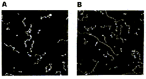
Single-stranded RNA
Single-stranded polyA (A, B) differs from double-stranded DNA mainly
in its persistence length. The persistence length of ss polyA is ca. 40
nm, while the persistence length of dsDNA under the same conditions is
ca. 80 nm. PolyA in water also protonates to form a very stiff double helix
with a persistence length of ca. 600 nm, (e.g., arrow in B) Images are
1 mm2. (From (1).)
Triple-stranded DNA
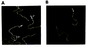
Features that appear to be triple-stranded DNA can be seen on poly(dA):poly(dT)
(A, B) and poly(dG):poly(dC). The putative triple-stranded DNA is ca. twice
as high as dsDNA. Images are 1 mm2. (From (1).)
Helix turns on dsDNA (2)
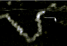
The best images of helix turns with tapping AFM show more detail than
images of helix turns with contact AFM (3, 4). These helix turns were imaged
in propanol with an unusually good tip and cannot be obtained reliably.
Scale bar is 10 nm. Shao's group can see helix turns on dsDNA much more
reproducibly but in less detail than the above image, using samples of
densely packed DNA on a positively charged lipid bilayer, imaged with contact
AFM (3). We have not yet been able to image such a sample with tapping
AFM to see if the helix turns can be resolved in more detail. The DNA in
propanol shows intriguing deformations of the double helix where the DNA
bends sharply that would be fascinating to image more. We believe that
only the major groove was detected, because the periodicities are 3-4 nm.
Densely packed DNA on lipid should be better for getting high resolution,
because there is less interaction between the tip and the DNA than with
isolated DNA molecules (5).
References:
1. Hansma, H. G., I. Revenko, K. Kim, and D. E. Laney. 1996. Atomic force microscopy of long and short double-stranded, single-stranded and triple-stranded nucleic acids. Nucleic Acids Res. 24:713-720.
2. Hansma, H. G., M. Bezanilla, D. L. Laney, R. L. Sinsheimer, and P. K. Hansma. 1995. Applications for Atomic Force Microscopy of DNA. Biophys. J. 68:1672-1677.
3. Mou, J., D. M. Czajkowsky, Y. Zhang, and Z. Shao. 1995. High-resolution atomic-force microscopy of DNA: the pitch of the double helix. FEBS Lett. 371:279-282.
4. Hansma, H. G., and P. K. Hansma. 1993. Potential applications of atomic force microscopy of DNA to the human genome project. Proc. SPIE - Int. Soc. Opt. Eng. (USA). 1891:66-70.
5. Hansma, H. G. 1995. Polysaccharide helices in the atomic force microscope. Biophys. J. 68:3-4.