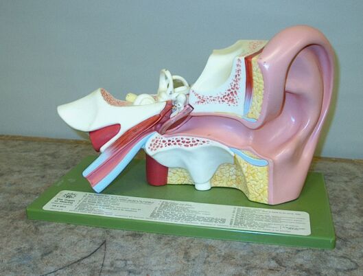
You can use this anatomical model to show the various parts of the human ear as you describe their functions.
Sound is a longitudinal mechanical wave created by a disturbance in an elastic medium, most commonly air. It is a series of alternating compressions, regions of higher-than-average pressure, and rarefactions, regions of lower-than-average pressure, which produce pressure fluctuations as the wave travels through the air. Our ears are sensitive to these pressure fluctuations, and by converting them to mechanical motion of various parts of the middle and inner ear, and then into a nerve signal that goes to the brain, they enable us to hear sound waves.
The ear has three sections: the outer ear, the middle ear and the inner ear.
The outer ear
The outer ear comprises the auricle (or pinna), which is the part on the outside of the head, essentially cartilage covered with skin, the (external) auditory canal, the tympanic membrane (or ear drum), and the tympanic ring. The auricle guides sound waves into the auditory canal. Its shape and orientation, in combination with the binaural hearing afforded by our having two ears, enhance our ability to determine the location of the source of a sound. The auditory canal guides the sound wave to the tympanic membrane, which is held in place by the tympanic ring, which is a ring of bone that surrounds it. Sound that reaches the ear drum sets it vibrating.
The middle ear
The middle ear comprises the tympanum, the cavity on the interior side of the tympanic membrane, in which sit three small bones, called the malleus (hammer), incus (anvil) and stapes (stirrup), so named because they resemble these objects. The flat end of the stirrup rests on the Fenestra vestibuli (or oval window). An opening at the bottom of the middle ear leads to the eustachian tube, which connects the middle ear to the nasopharynx. This allows for equalization of the pressure on both sides of the tympanic membrane. When set vibrating by an incoming sound wave, the tympanic membrane in turn sets the malleus vibrating, which sets the incus vibrating, which sets the stapes vibrating.
The part of the model that comprises the tympanic membrane, malleus and incus, is removable to make these parts more visible.
The inner ear
The inner ear comprises the vestibule, which is on the interior side of the oval window, three semicircular canals (lateral, anterior and posterior), which sit above the vestibule, the cochlea (from the Latin for “snail” or “snail shell,” from the Greek kochlias, from kochlos, “snail,” because it resembles a snail shell) and the auditory nerve, which transports the signal from the cochlea to the auditory cortex in the brain. The semicircular canals are fluid-filled ducts, roughly mutually orthogonal to each other, inside which cilia (hair cells) detect motion of the fluid. This provides us with our sense of balance. Vibrations are transferred from the stapes through the oval window, to the fluid that fills the vestibule and cochlea. The cochlea comprises three chambers, all coiled side by side around a common axis. The central chamber contains the organ of Corti, which contains rows of cilia, which detect vibrations in the cochlear fluid, and transmit the resulting signal to the auditory nerve, which carries it to the brain.
The part of the model that comprises the stapes, vestibule, semicircular canals, cochlea and a section of the auditory nerve, is removable to make these parts more visible.
The ear is exquisitely sensitive. At a reference frequency of 1,000 Hz, it can detect pressure fluctuations as small as about 2 × 10⁻⁵ Pa (above and below atmospheric pressure). On average, atmospheric pressure at sea level is 1.013 × 10⁵ Pa, so this represents a relative change in pressure of about ±1 part in 5,000,000,000. The loudest sound that the ear can receive without pain corresponds to a pressure fluctuation of about 30 Pa, or about ±1 part in 3,400, or over 1,000,000 times greater than the smallest detectable pressure fluctuation. This pressure fluctuation (30 Pa), at 1,000 Hz, corresponds to an amplitude, or maximum displacement, of about 1.2 × 10⁻⁵ m (~12 μm). For the softest detectable sound (pressure fluctuation of 2 × 10⁻⁵ Pa), at 1,000 Hz, the maximum displacement is about 10-11 m, or about 0.1 Å, which is on the order of 1/10th the diameter of an atom! For another reference, the wavelength of yellow light is about 600 nm, or 6 × 10⁻⁷ m (0.6 μm), or 60,000 times greater than this displacement! This sensitivity to pressure fluctuations over a range of over six orders of magnitude is quite impressive.
The intensity of a wave is defined as the time average rate at which energy is transported by the wave per unit area across a surface perpendicular to the direction in which the wave propagates. Its units are W/m2. The intensity is proportional to the square of the amplitude of the wave, and thus the square of the pressure fluctuation. The smallest audible pressure fluctuation, about 2 × 10⁻⁵ Pa, corresponds to a power of 10-12 W/m2, and the pressure fluctuation at the threshold of pain, 30 Pa, corresponds to a power of 1 W/m2. We see that the six-order-of-magnitude range of sensitivity to the pressure fluctuation corresponds to a range of twelve orders of magnitude in power of the sound waves that the ear can detect.
As you might imagine, the ear’s response to sound intensity over this wide range is not linear, but rather it is logarithmic. We perceive changes in the intensity of sounds by equal ratios as equal changes in loudness. That is, if we are exposed to sounds whose intensities differ by a common multiplier, we hear the changes in intensity from one to the next as equal changes in loudness. In order for one sound to appear twice as loud as another, it must have 10 times the intensity. (See demonstration 44.09 -- Sound level meter.)
The nominal range of frequencies over which the ear responds is from 20 to 20,000 Hz. Sound waves with frequencies below 20 Hz are referred to as infrasonic, and those with frequencies above 20,000 Hz are said to be ultrasonic. As is the case for intensity, the ear’s response to frequency is also nonlinear. We hear equal frequency ratios as equal pitch intervals. For example, any two pitches that have a frequency ratio of 2:1 sound an octave apart. Any two pitches whose frequencies are in a ratio of 3:2 sound a perfect fifth apart. Any two pitches whose frequencies are in a ratio of 4:3 sound a perfect fourth apart. Other intervals with their ratios are major third, 5:4; minor third, 6:5; major sixth, 5:3; minor sixth, 8:5; major seventh, 15:8; minor seventh, 16:9; major second, 9:8 and minor second, 16:15.
References:
1) Wikipedia entry on the Cochlea (The anatomy and function of the cochlea; a detailed description with excellent diagrams, and related links.)
2) https://www.verywellhealth.com/ear-anatomy-4843989
3) https://www.verywellhealth.com/semicircular-canals-anatomy-of-the-ear-1191868
4) https://www.verywellhealth.com/cochlea-anatomy-5069393
5) Berg, Richard E. and Stork, David G. The Physics of Sound (Englewood Cliffs New Jersey: Prentice-Hall, Inc., 1982) pp.140-145.
6) Young, Hugh D. and Geller, Robert M. Sears & Zemansky’s College Physics (San Francisco: Addison Wesley, 2007) pp. 385-6.
7) Sears, Francis W. and Zemansky, Mark W. College Physics, Third Edition (Reading, Massachusetts: Addison-Wesley, 1960) pp. 431-7.
8) http://hyperphysics.phy-astr.gsu.edu/hbase/Sound/loud.html
9) Apel, Willi. Harvard Dictionary of Music (Cambridge, Massachusetts: Harvard University Press, 1955) pp. 359-363 (Intervals, and Intervals, Calculation of).