This demonstration is the result of a collaborative effort by Robert Geller, Louis Grace and Cord Dorcey.
Various organisms are sensitive to the earth’s magnetic field, and can detect its orientation and use it for navigation or other purposes. The simplest of these are various single-celled animals. These organisms synthesize within their cell membrane tiny crystals of magnetite, which they incorporate into organelles called magnetosomes. It is these organelles that allow these animals to detect the earth’s magnetic field, and thus give them the capability of magnetotaxis, that is, translational or orientational motion in response to an external magnetic field. Hence their designation as magnetotactic. These bacteria thrive in environments with limited amounts of oxygen. With either too much or too little oxygen, they die. One finds them in stratified environments, either aquatic areas or in soil, where they use the earth’s magnetic field to direct them downwards until they reach a region where the oxygen level is optimal. This means that they follow the magnetic field lines into the earth, so that in the northern hemisphere the bacteria are attracted to geomagnetic north. (See demonstration 68.09 -- dip needle.) There is some question regarding the details of this behavior, since someone recently found, in the northern hemisphere, a bacterium that appears to be attracted to geomagnetic south. (See Magnetic Misfits: South Seeking Bacteria in the Northern Hemisphere.) In any case, as best anyone can tell, these bacteria use the earth’s magnetic field to navigate towards a region where the environment is favorable.
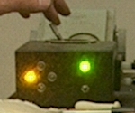
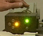
Our magnetotactic bacteria are monococci, that is, bacteria consisting of a single, roughly spherical unit. They also have a flagellum at one spot to provide locomotion. Our first samples came from the Goleta slough, one from a point near Walnut Lane, and another from near Puente Drive. We also used soil from outside the Broida lecture hall building. We store the samples in mason jars with their metal lids loosely set. To harvest the bacteria, we tighten the lid, shake the jar to distribute the contents, then place a magnet next to the side of the jar, about three or four centimeters below the surface of the water. Since geomagnetic north is actually a south magnetic pole, we place the magnet so that its south pole faces the jar. The bacteria swim toward a spot near the magnet, so that after some time, if we shine a flashlight at the wall of the jar, we can see a white deposit where the bacteria have gathered. We reach in with a pipette, pull some fluid near that spot, then place a small drop on a microscope cover glass. In the original version of this demonstration, we flipped this cover glass over, so the drop hung from the bottom, then set it on an o-ring that sat on a microscope slide. We then placed the slide onto the microscope stage (in a manipulator) and looked for the front edge of the drop, then placed a magnet near the slide, south pole facing the slide, north facing away, and watched as the bacteria gathered against the drop edge. Not only does the edge provide a good stop for the bacteria, but since the drop is thinnest there, problems caused by the narrow depth of field at 1,000X magnification are not as bad as they would be elsewhere in the drop. (You can find an example of this method at this link. We do not use the “racetrack” apparatus described in the paper. The liquid drawn through the tip of a pipette placed near the spot where the bacteria gather on the jar wall, contains enough bacteria to make a healthy crowd as viewed under the microscope.) While this method is still available, we now use a plastic plate with twelve cylindrical rare earth magnets, 1/16 inch in diameter and 1/8 in long, placed in a circle, north pole up, embedded in it, with a hose washer as a spacer on which we place the cover slip with the hanging drop. The photograph below shows this rig:
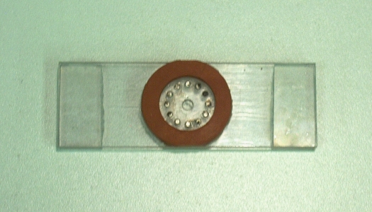
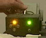
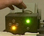
The bacteria swim away from the north poles of the ring of magnets, and they gather at the center, against the bottom surface of the cover slip. Placed at right angles near the microscope stage are two electromagnets, current for which is provided by the power supply in the black case sitting on the middle shelf of the cart (with the red LED lit at left). The unit sitting to the left of the microscope in the photograph (with the green LED lit at right), allows you to control the current through the electromagnets by means of a joystick. For either presentation method, with the drop hanging below the cover glass, we need not worry about the microscope objective touching the drop. A video camera placed immediately above the ocular of the microscope captures the image. (The camera is oriented so as to erect the normally inverted microscope image.) A video monitor on the bottom shelf of the cart allows us to display the image during setup. A cable from the monitor connects to the video port of a data projector in the lecture hall so the class can see the image.
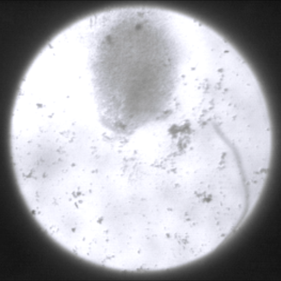
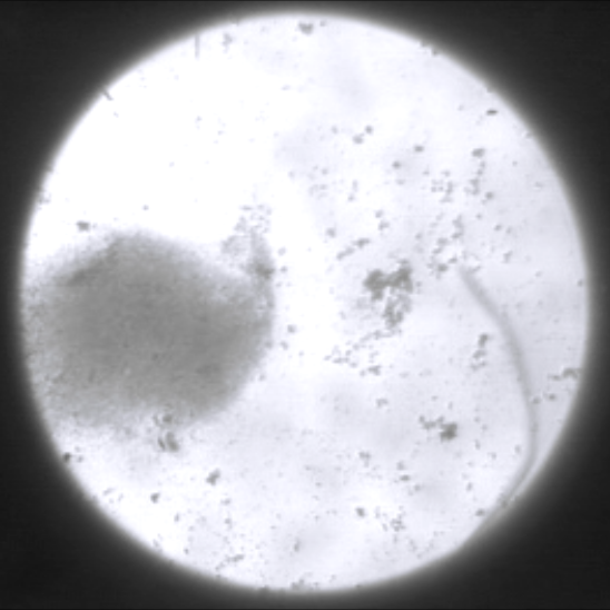
The photograph below shows a large crowd of bacteria gathered at the center (cover glass placed on the rig shown above, no current in the electromagnets, so no external magnetic field superimposed on that from the ring of rare earth magnets):
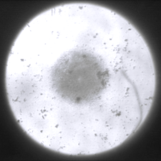
For the photograph above, the magnification is 100X. Normally, it might be desirable to have a smaller group, with which it is possible to use higher magnification. It is, however, somewhat difficult to control the number of bacteria in the sample. (The microscope also offers magnifications of 400X and 1000X. At 400X, the group of bacteria in the photograph above would more than fill the field.)
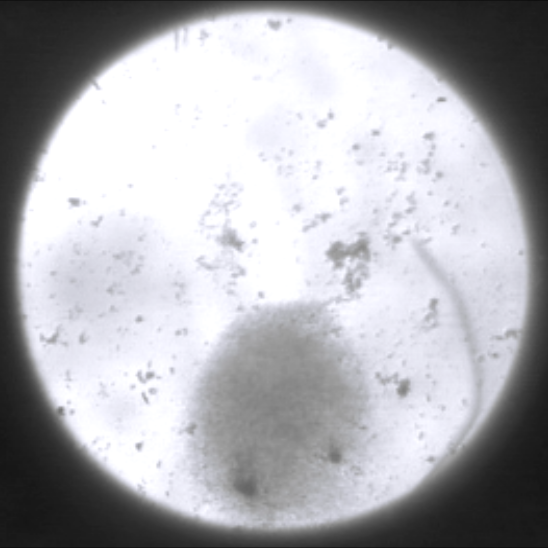
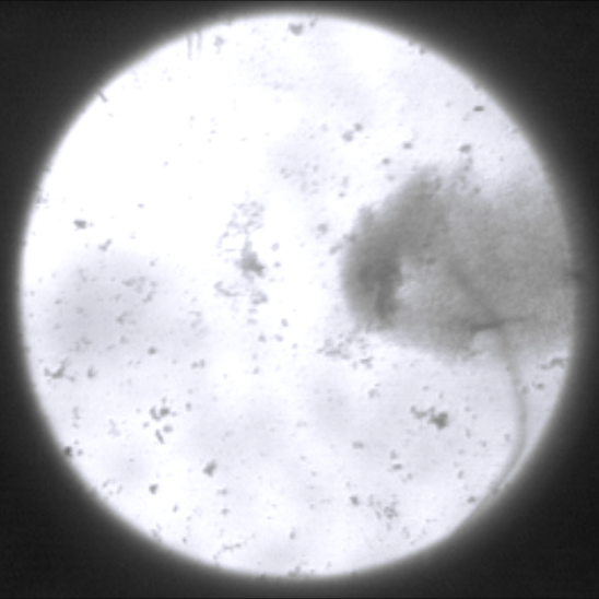
The photographs below show the motion of the bacteria when an external magnetic field is imposed by means of the electromagnets:
the microscope, the bacteria swim downward.
faces the microscope, the bacteria swim rightward.You can, of course, move the joystick in any direction, and thereby send current to both electromagnets at the same time, in varying proportions. In this way, you can make the bacteria swim linearly in any direction, or lead them around in circles, etc.
Below are photographs from the original arrangement. The first photograph shows the bacteria that have gathered at the drop edge after it has sat in the field of a cow magnet for a short while. The line near the top of the field, below which you can see the bacteria, is the edge of the drop. Magnification is 1000X.

When we remove the magnet and turn it around, the bacteria swim away:

By moving the magnet in different ways, you can make the bacteria swim toward the drop edge, away from the drop edge or across the field.
References:
1) Lins, Ulysses; Freitas, Flavia; Keim, Carolina Neumann; de Barros, Henrique Lins; Esquivel, Darci Motta S.; Farina, Marcos. Brazilian Journal of Microbiology (2003) 34, 111-116.