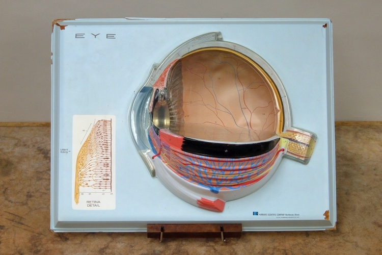

You can use this model to show the various parts of the human eye as you describe their functions.
We see the objects around us by means of the light that they reflect or, in some cases, emit. The electromagnetic spectrum spans a wavelength range from on the order of 1,000 m (the longest-wavelength AM radio signals are about 560 m at a frequency of 540 kHz) to on the order of about 10-12 m (gamma rays at a frequency of about 3 × 10²⁰ Hz.) Our eyes are sensitive to light in the wavelength range from approximately 700 nm to 400 nm (~4.3 × 10¹⁴ Hz to ~7.5 × 10¹⁴ Hz). This band of the electromagnetic spectrum is therefore called the visible spectrum, and light within this range is known as visible light. The color of the light at the end of the spectrum near 700 nm is red, and at the end near 400 nm it is blue. Because the energy of the light increases from the red end of the spectrum to the blue end, light having a wavelength of ~700 nm to ~1 mm is known as infrared, and light having a wavelength of ~400 nm to ~10 nm is called ultraviolet.
Light enters the eye through a curved transparent layer of tissue called the cornea, behind which is a small chamber called the anterior chamber, which is filled with a fluid called the aqueous humor. On the other side of the anterior chamber is the iris, whose muscles are responsible for the contraction and expansion of the aperture at its center, called the pupil. Behind the iris is the lens, sometimes called the crystalline lens, which is ringed by the ciliary muscle. Behind the lens, filling the rest of the space within the eyeball, is a fluid called the vitreous humor. Lining the majority of the inner surface of the eyball in this region is the retina, which contains the rod cells and cone cells that detect the incoming light, and also a layer of nerves that process the resulting signals and send them to the brain via the optic nerve.
The cornea and lens focus the light that enters the eye, to form a real, inverted image on the retina. As noted above, the cornea is curved. It has an index of refraction of about 1.376, and it performs most of the refraction of the incoming light. The lens is made up of fine layers, and varies in index of refraction from about 1.406 in the center to about 1.386 toward the edge. The index of refraction of the aqueous humor (between the cornea and the lens) is about 1.336, close to that of water (1.333), and the vitreous humor has an index of refraction of about 1.337. For viewing objects at a distance, the ciliary muscle is relaxed, and the eye focuses at infinity. To focus images from objects that are close to the eye, the ciliary muscle contracts, which allows the lens to become more greatly curved (it bulges; the radii of curvature of both surfaces decrease), thus shortening its focal length. This is called accommodation.
The iris, which as noted above, sits between the anterior chamber and the lens, regulates the amount of light that enters the eye. In low-light situations, the radial muscles of the iris contract while the circular muscles relax, and the pupil expands. Under bright light, the circular muscles contract while the radial muscles relax, and the pupil contracts. This process is called adaptation. The range over which the diameter of the pupil varies is only about a factor of four, for a 16-fold range in area, and hence in the amount of light it allows into the eye. The variation in brightness, however, from an outdoor scene in bright sunlight, to a dimly lit room, for example, can be several orders of magnitude. The retina is able to adapt to large differences in brightness. Together, the adaptation of the iris and the adaptation of the retina allow the eye to adapt over a wide range of light intensity.
The retina, as noted above, lines the inside of the eyeball over most of the surface in the region that holds the vitreous humor. Behind the retina is a layer of cells pigmented with melanin. The retina consists of three layers, separated by synapses between the axons and dendrites of the cells in adjacent layers. The outermost layer is the layer of rods and cones, which detect light and convert it to a nerve signal. Lining the layer of rods and cones is a layer that contains three types of nerve cells: bipolar cells, horizontal cells and amacrine cells. (Most of the cells in this layer are bipolar cells.) Bipolar cells receive signals from the rods and cones, and many of them communicate directly with cells in the next layer, called ganglion cells. Horizontal cells connect rods or cones with bipolar cells by relatively long connections that run parallel to the layers of the retina. Amacrine cells connect bipolar cells with ganglion cells in similar fashion. The innermost layer of the retina is the layer of ganglion cells, with the layer of nerve fibers that connect them to the optic nerve.
The rods are extremely light sensitive. The fully dark-adapted eye can see a brief flash of light that is so dim that no single receptor can have received more than one photon. According to calculations, in order for the eye to see a flash, around six closely spaced rods must be stimulated, each by a single photon, within a short time. The light-absorbing pigment in the rods is rhodopsin, whose absorption maximum is in the green region of the visible spectrum. Rhodopsin thus reflects in the red and blue regions of the spectrum, which makes it appear purple. For this reason it is also called visual purple.
There are three types of cone cell. Each type has a pigment whose absorption peaks in a different region of the visible spectrum from where those of the other two types peak. The absorption peaks lie at about 445 nm, about 535 nm and about 575 nm. Because of where these wavelengths fall in the spectrum, by convention, these cones and their pigments are referred to as blue, green and red, respectively. It bears noting that this is inaccurate in that colors of these wavelengths would not appear blue, green or red, and if we stimulated only one type of cone, we might see green, but blue would probably appear violet, and red would probably appear yellowish-red. Also, whereas rhodopsin is named for the color of light that it reflects, the cone pigments are named for the colors that they absorb.
The retina is, in a sense, inside out, in that light must pass through the layer of ganglion cells and the layer of bipolar cells before it can reach the layer of rods and cones. One possible reason for this involves the function of the melanin-pigmented layer that lies behind the retina. This layer absorbs light that has passed through the retina, to prevent its scattering around the inside of the eye. (This is similar to the action of the black coating inside a camera.) The cells in this layer also chemically restore the pigment in the receptors after it has been bleached by light. To perform these functions, the pigmented layer must be close to the receptors. If the rods and cones were at the front of the retina, the pigmented cells would have to sit between the layer of rods and cones, and the layer of bipolar cells. The presence of all the dendrites, axons and synapses in this region would make this quite difficult. Fortunately, the layers in which the bipolar cells and ganglion cells sit are essentially transparent and probably do not blur the image much. In the small region at the center of the retina, however, where visual acuity is greatest, these layers are displaced to form a ring where the retina is thicker than elsewhere, to expose the cones in the center. The shallow depression in which these cones lie is called the fovea. This area, about one millimeter in diameter, is where the cones are most densely packed, and contains only cones. Cones are distributed throughout the retina, but are greatly outnumbered by rods.
Rods, which as noted above are quite sensitive to light, enable us to see in dim light, but saturate in bright light. Cones do not respond in dim light, but enable us to see fine detail, and they provide our color vision. The range of light intensities over which the eye responds is quite impressive. As noted above, the rods allow us to see light that is very dim, probably on the order of a few μcd/m2. The maximum intensity the eye can see without discomfort is perhaps around 105 cd/m2. (The candela is defined as the luminous intensity, in a given direction, of a monochromatic source emitting at a frequency of 5.40 × 10¹⁴ Hz, that has a radiant intensity in that direction of 1/683 watt per steradian.) Thus, the eye can respond to light over a range of intensities that spans over 10 orders of magnitude. As noted above, in very dim light, rods respond but cones do not respond, and in very bright light, the rods are saturated and the cones respond. At intermediate light levels, both rods and cones respond. This leads to three types of vision. At light levels below about 0.003 cd/m2, only the rods respond, and there is no sensation of color. Vision in this range is said to be scotopic. At light levels above about 3 cd/m2, only the cones respond. Vision in this range is said to be photopic. At light levels between about 0.003 cd/m2 and about 3 cd/m2, both rods and cones respond. Vision in this range is said to be mesopic.
Various optical conditions of the eye
In a normal eye, light enters and is focused by the cornea and lens to form a real, inverted image on the retina. It is not uncommon, however, for defects in the eye to cause the image not to focus at the retina.
In myopia, or nearsightedness, because of defects in the cornea, lens or shape of the eyeball, or some combination of these, light entering the eye focuses in front of the retina. A diverging lens of the appropriate (negative) focal length, placed in front of the eye, causes the incoming light rays to diverge, thus moving the image back so that it focuses on the retina.
In hyperopia, or farsightedness, the lens and cornea do not cause the incoming light rays to converge enough, and the image focuses behind the retina. A converging lens of the appropriate (positive) focal length, placed in front of the eye, causes the incoming light rays to converge to a greater extent, and thus focus at the retina.
As noted above, for viewing distant objects, the eye focuses at infinity, and for viewing objects that are close, the ciliary muscles contract to increase the curvature, and thus shorten the focal length, of the lens. The shortest object distance for which accommodation can bring the image into focus is called the near point. Because the flexibility of the lens decreases with age, the distance to the near point increases with age. It ranges, on average, from about 7 cm for a 10-year-old, to about 22 cm for a 40-year-old, and continues to increase with age. This recession of the near point with age is called presbyopia. As the near point recedes, it becomes more and more difficult to perform tasks that require close focus, such as reading. For people with otherwise good vision, this results in a need for reading glasses. For people who require corrective lenses to achieve normal distance vision, it results in a need for bifocals, which have the correction for distance vision in the top portion of the lens and the correction for near vision in the bottom portion, or progressive lenses, which gradually change from the correction for distance vision in the top portion of the lens to the correction for near vision in the bottom portion of the lens.
Astigmatism is a nonuniformity in the curvature of the cornea, that causes parts of an image that lie along one particular axis to focus differently from those that lie along the axis that is perpendicular to it. To correct for this, one places a cylindrical lens in front of the eye, with its axis of curvature aligned with the axis along which the curvature of the cornea deviates from the curvature in other directions. Whereas a spherical lens focuses light in all planes, since the cylindrical lens is curved in only one plane, it affects the focus of the light in only that plane. Such a correction can be added to a lens that corrects either for myopia or for hyperopia.
The lenses used to correct for myopia or hyperopia are spherical lenses that have negative or positive focal lengths, respectively. The optical power, or strength, of such lenses is quoted in units called diopters, which equal the inverse focal length of the lens in meters. For example, a positive (converging) lens whose focal length is 2.0 meters has a power of +0.50 diopters, and a negative (diverging) lens with a 2.0-meter focal length has a power of -0.50 diopters. A positive lens with a focal length of 0.50 meters has a power of +2.0 diopters, and a negative lens whose focal length is 0.50 meters has a power of -2.0 diopters. On a prescription for eyeglasses, such corrections are listed as spherical corrections, in diopters. If a correction for astigmatism is also necessary, it is listed as a cylindrical correction in diopters, with the angle at which the cylinder axis lies. (90° lies on the vertical axis, and 180° lies on the horizontal axis).
References:
1) http://hyperphysics.phy-astr.gsu.edu/hbase/vision/eyescal.html#c1
2) http://hyperphysics.phy-astr.gsu.edu/hbase/vision/colcon.html
3) https://www.engineeringtoolbox.com/refractive-index-d_1264.html
4) Young, Hugh D. and Geller, Robert M. Sears & Zemansky’s College Physics (San Francisco: Addison Wesley, 2007) pp. 841-46.
5) Feynman, Richard P.; Leighton, Robert B. and Sands, Matthew. The Feynman Lectures on Physics, Volume I (Menlo Park, California: Addison-Wesley Publishing Company, 1963) pp. 35-1,2; 36-3,4.
6) Hubel, David H. Eye, brain, and vision (Scientific American Library series; #22) (New York: Scientific American Library, distributed by W.H. Freeman and Company, 1988) pp. 34-9, 49, 162-4.
7) Sears, Francis W. and Zemansky, Mark W. College Physics, Third Edition (Reading, Massachusetts: Addison-Wesley, 1960) pp. 848-853.
8) https://www.ecse.rpi.edu/~schubert/Light-Emitting-Diodes-dot-org/Sample-Chapter.pdf
9) https://www.telescope-optics.net/eye_intensity_response.htm
10) https://www.olympus-lifescience.com/en/microscope-resource/primer/lightandcolor/humanvisionintro/
11) https://www.nist.gov/si-redefinition/candela
12) https://www.aao.org/eye-health/diseases/what-do-astigmatism-measurements-mean