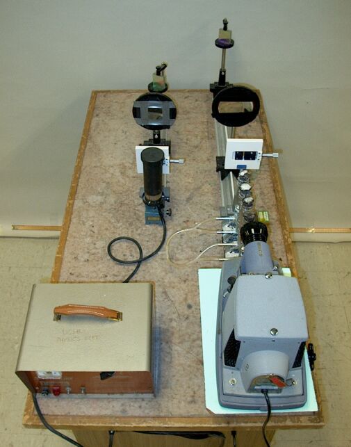
Light from a projector (bottom right) passes through a slit, whose image is focused onto a grating. The diffracted light appears as a continuous spectrum on a portable screen (not shown). When you light the four Meker burners, the flames vaporize table salt (sodium chloride) sitting on screens placed just above the burners. The light from the projector must pass through the resulting vapors on its way to the slit. Though the flames appear orange from emission by excited (neutral) sodium atoms, many more are in the ground state, and they absorb light from the projector at the same wavelengths. This gives rise to two closely-spaced dark lines in the orange region of the continuous spectrum. A sodium lamp, placed behind the slit on the shorter optical rail on the table, allows you to display the sodium emission lines. The rails and gratings are aligned so that the emission lines appear below the absorption lines in the continuous spectrum, to illustrate that the emission and absorption occur at the same wavelengths. On the same table, but not in the photograph above, is a second power supply connected to a mercury lamp. This lamp produces a series of emission lines ranging from the yellow to the ultraviolet. Again, the rail and grating are aligned with the longer rail so that the lines appear below their respective positions within the continuous spectrum. A paddle (not shown), one surface of which is coated with a fluorescent paint, and the other side of which has one half coated with fluorescent paint and the other with non-fluorescent paint, allows you to show that there are emission lines outside the range of the visible spectrum (in the ultraviolet).
Demonstration 84.06 -- Laser beam diffracted through various slits, shows diffraction by a single slit, the combination of diffraction and interference by double slits, and how the pattern produced by diffraction and interference changes as one increases the number of slits from two to three, four and five (all equally wide and equally spaced). A pair of slits produces a diffraction pattern that has within each diffraction maximum a series of interference maxima and minima, or fringes. As one increases the number of slits, the variation in intensity caused by diffraction decreases, and the intensity of the interference maxima comes closer to being uniform across the pattern. The behavior begins to resemble that of a diffraction grating, which is a device made of many closely-spaced parallel slits (or grooves). Depending on the method of production, these grooves are either etched or deposited on some kind of substrate. If the substrate is transparent (e.g., plastic or glass), the incident light passes through, and the grating is a transmission grating. If the substrate is reflective or is coated with a reflective material (e.g., aluminum or gold), then incident light is reflected, and the grating is a reflection grating, as are the gratings used in this demonstration. Demonstration 84.18 -- Laser diffracted by reflection gratings, shows the patterns produced by several diffraction gratings, each of which has a different line spacing. (The gratings in this demonstration have a spacing of 600 lines per millimeter.)
As for the two-slit system, the locations of the (principal) interference maxima on a screen on which light coming from the grating is projected, are determined by the slit spacing and the wavelength of the light, and are given by the equation
d sin θ = mλ, where m = 0, 1, 2, . . .
d is the slit spacing, θ is the angular displacement of the particular maximum from the center of the pattern, and λ is the wavelength. m is called the order number. The central maximum is the zeroth order. Going outward from the center, located to either side are the first order, second order, etc. As for double slits, if D, the distance from the slits to the screen, ≫ y, the distance of a point on the screen from the center of the screen, then sin θ ≈ tan θ ≈ y/D, and dy/D = mλ (or y = mλD/d; m = 1, 2, 3, . . .). This approximation does not hold for orders that appear far from the center of the screen.
We see from the above that the location of each maximum on the screen (except for the zeroth-order maximum) is wavelength dependent. With the appropriate setup, then, one can use a diffraction grating to measure the wavelength(s) of light emitted by a source. One can build an instrument in which a narrow beam of light from the source is diffracted by a grating to fall on a piece of film, a detector array or a screen. Either by means of a built-in scale calibrated in wavelength, or by comparison to the locations of known lines from a reference source, one can measure the wavelengths for the various lines that appear on the detector. One can also sample a narrow beam of the diffracted light with a single detector, turn the grating slowly over a range of angles, and observe the resulting maxima in intensity that occur. In such an instrument, the drive that turns the grating is typically calibrated in units of wavelength, typically nanometers or ångstroms, and one obtains a plot of intensity vs. wavelength. (It is common to use a reference source to check the calibration of the wavelength scale.) The various lines one observes from a source make up a spectrum, and an instrument that allows one to measure the wavelengths of these lines is called a spectrometer.
The two systems used in this demonstration are not really spectrometers, but they illustrate how such an instrument works. For the large optics rail with the burners, the projector is the light source, and for the small optical rail, either the sodium lamp or the mercury lamp is the light source. The light from each source is diffracted by a reflection grating, and light of different wavelengths appears at different places on the screen, according to its wavelength, light of longer wavelengths displaced from the location of the zeroth order by greater distances than light of shorter wavelengths.
The projector lamp emits broadly in the visible spectrum, with some emission in the ultraviolet and a significant amount in the infrared. We see this emission as white light. Since the lamp filament emits essentially blackbody radiation, the spectrum of wavelengths at which it emits light is continuous, and the light is spread out over a range of angles. Light in the visible region thus appears as the familiar rainbow pattern. When you light the four Meker burners that sit between the projector and the slit, the flames heat table salt (sodium chloride) sitting on screens placed just above the burners. This vaporizes some of the salt, and the light from the projector must pass through the vapors on its way to the slit. (When the sodium chloride vaporizes, the electrons stay with the sodium atoms, so the sodium vapor consists of neutral sodium atoms, and the chlorine is released as Cl2.) Since the sodium vapor is hot, some of the atoms are in an excited state, and when they relax to the ground state they emit orange light. Most of the sodium atoms, however, are in the ground state, and when the light from the projector lamp passes through the flames, these sodium atoms absorb light and make the transition from the ground state to an excited state. The light that they absorb is the same wavelength as the light emitted by the excited atoms, and the absorption results in a dark line in the orange region of the spectrum produced by the projector lamp. If you place the sodium lamp in front of the smaller optical rail, you can show the sodium emission line. The grating on the small rail is aimed so that this emission line appears below the dark line in the continuous spectrum.
The orange emission line is actually a doublet, the sodium D lines, at 5,890.0 Å and 5,895.9 Å, and is due to transitions from the 2P3/2 and the 2P1/2 excited states, respectively, to the 2S1/2 ground state. These transitions occur when the valence electron in an excited sodium atom relaxes from a 3p orbital to the 2s orbital. As the light from the projector passes through the flames, some of it is absorbed when it excites sodium atoms from the 2S1/2 ground state to either the 2P3/2 or the 2P1/2 excited state by exciting an electron from the 2s orbital to a 3p orbital. (The dark absorption line is also a doublet.)
The symbols 2P3/2, 2P1/2 and 2S1/2, above, are called term symbols. The left superscript denotes the spin multiplicity, 2S + 1, where S is the total electron spin for the state. The italic capital letter, S, P, D, F, etc., denotes the total orbital angular momentum of the valence electron(s), 0, 1, 2, 3, etc. The right subscript denotes J, the vector sum of the spin and orbital angular momenta, L + S. (J has the range L + S, L + S - 1, L + S - 2, . . . , |L - S|.)
If you place the mercury lamp in front of the small rail, you can show the emission spectrum of mercury. The mercury lamp produces a series of emission lines ranging from the yellow to the ultraviolet. Again, the rail and grating are aligned so that the lines appear below their respective positions in the continuous spectrum. A paddle (not shown), one surface of which is coated with a fluorescent paint, and the other side of which has one half coated with fluorescent paint and the other with non-fluorescent paint, allows you to show that there are emission lines outside the range of the visible spectrum. These are higher in energy than violet, i.e., they are in the ultraviolet. You can also get a volunteer who has a white or light-colored freshly-laundered shirt. The UV shifters (optical brighteners) in the laundry detergent fluoresce when exposed to UV, and the UV lines are thus visible on the student’s shirt. The fluorescent molecules in the coating on the paddle, or in the brighteners in laundry detergent, absorb the UV light and become electronically excited. They then undergo a radiationless transition to a lower-energy state, from which they relax to the ground state and emit a photon in the process. The difference in energy between this intermediate state and the ground state corresponds to that of a photon whose wavelength is in the visible spectrum. Such molecules typically have available multiple levels in the excited state, and can undergo transitions to multiple levels in the intermediate state. Because of this, and also because of the short lifetime of the intermediate state, both the absorption and fluorescence emission tend to be broadband. Thus, all of the UV lines coming from the mercury lamp appear as white or blue-white lines on the fluorescent paddle.
The yellow, green, blue and violet lines, and one at the edge of the violet and ultraviolet, from the mercury lamp are readily identifiable. The pair of yellow lines appears at 5,790.66 Å and 5,769.60 Å, and is due to transitions from the 1D2 state to the 1P1 state (6d to 6p), and the 3D2 state to the 1P1 state (6d to 6p), respectively. The second of these is spin forbidden, because in making the transition the electron undergoes a change of spin. Normally, the lines arising from such transitions do not appear, or are very weak. Because of the high charge on the mercury nucleus, however, the coupling between the orbital and spin angular momenta is great enough that this selection rule breaks down. As a result, some lines that would otherwise be absent or very weak, appear rather strongly.
The green line, at 5,460.74 Å, is from the transition from the 3S1 state to the 3P2 state (7s to 6p), the weak blue line, at 4,916.04 Å, is from the transition from the 1S0 state to the 1P1 state (8s to 6p), and the violet line, at 4,358.34 Å, is from the transition from the 3S1 state to the 3P1 state (7s to 6p). The line at 4,046.56 Å, which may not be visible without the fluorescent paddle, is from the transition from the 3S1 state to the 3P0 state (7s to 6p).
Three or four other lines are observable. Based on comparison with other mercury spectra, and on a fit of the wavelength vs. line position, the best candidates for these lines are those at 3,906.40 Å, 3,650.15 Å, 3,341.48 Å and 3,125.66 Å. (The one at 3,906.40 Å is weak.) These lines arise from the following transitions, respectively: 1D2 to 1P1 (8d to 6p), 3D3 to 3P2 (6d to 6p), 3S1 to 3P2 (8s to 6p) and 3D2 to 3P2 (6d to 6p).
In all of the transitions above, the electron is going from one orbital in the upper state to a different orbital in the lower state. These transitions are electric dipole transitions, which means that the two states between which the transition occurs cannot have the same symmetry. (The electric dipole operator is odd, so if the wavefunctions of the two states have the same symmetry, their integral with the operator vanishes.) This means that an electron in a particular orbital (s, p, d, f, etc.) in one level cannot go to the same orbital in a different level; such a transition is said to be symmetry forbidden. In making a transition, the electron must undergo a change in orbital angular momentum of one unit; that is, Δl = ±1. An s electron can go to a p orbital, a p electron can go to either an s orbital or a d orbital, a d electron can go to either a p orbital or an f orbital, etc. In addition, there is a constraint regarding J, which is ΔJ = 0, ±1; no transition between two states if both have J = 0.
References:
1) Halliday, David and Resnick, Robert. Physics, Part Two, Third Edition (New York: John Wiley and Sons, 1977), pp. 1047-8, 1051-2.
2) http://hyperphysics.phy-astr.gsu.edu/hbase/phyopt/grating.html#c1 and links to related material.
3) Herzberg, Gerhard, Spinks, J. W. T. translator. Atomic Spectra and Atomic Structure (New York: Dover Publications, 1945) pp.74, 116, 160 (Sodium D lines), 153 (Selection rules).
4) Specification sheet for an Osram high-pressure mercury lamp, with partial spectrum: http://www.lamptech.co.uk/Spec%20Sheets/D%20SP%20Osram%20Spectral%20Hg.htm
5) NIST mercury spectrum, strong lines (Hg I is the neutral atom; Hg II is the singly-charged ion (Hg+)): http://physics.nist.gov/PhysRefData/Handbook/Tables/mercurytable2.htm
6) NIST mercury spectrum, persistent lines: http://physics.nist.gov/PhysRefData/Handbook/Tables/mercurytable3.htm
7) Herzberg, Gerhard, Spinks, J. W. T. translator. Atomic Spectra and Atomic Structure (New York: Dover Publications, 1945) p.6, Fig. 5. Spectrum of a Mercury-Vapor Lamp, pp. 154-155, p. 202, Fig. 75. Energy Level Diagram of Hg I, from Grotrian, W., Graphische Darstellung der Spektren von Atomen und Ionen mit ein, zwei und drei Valenzelektronen (Springer, Berlin, 1928).